Non-Carious Cervical Lesions (NCCL): Causes and Contributing Factors
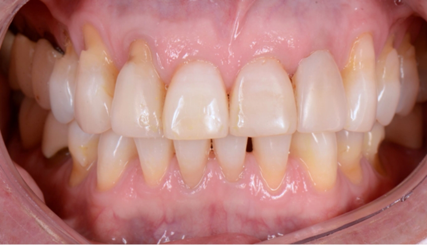
A non-carious cervical lesion (NCCL) is defined as the loss of tooth structure at the cementoenamel junction (CEJ) under pathological conditions. These lesions are often called abfractions, but this term is a misnomer, and I’ll soon explain why. These lesions most frequently occur on the facial surfaces of teeth, but it’s not entirely impossible to spot them on the lingual or interproximal surface. Epidemiology studies report that this phenomenon most frequently occurs in premolars (1, 2). Patients typically report hypersensitivity because of enamel rod corrosion and dentin tubule exposure.
It’s important to note that the loss of tooth structure associated with NCCLs is not attributed to caries (hence non-carious cervical lesion). There is often speculation about the cause of NCCLs. Current advances from the last decade have helped shed light on the etiology of NCCLs and provide management strategies. I would also like to propose that we, as a community, refrain from using the term “abfraction” to describe non-carious cervical lesions exclusively. Abfraction is one of the mechanisms that may cause an NCCL, but the lesion may occur from other forces.
How Abfraction Theory Ignores the Bigger Picture

Etiology
Today, there is extensive evidence that NCCLs are multifactorial and not necessarily caused by a single variable. NCCLS may be caused by the synergistic action of erosion, friction (abrasion), and possibly occlusal stress (abfraction). Below, I will discuss those different factors and explain them.
Erosion
While dietary acid is a source of erosion, it is certainly not the only one. Intrinsic acids (gastric acid) are a common source of erosion. Patients should be screened for a history of GERD, bulimia nervosa, and hiatal hernias. Interestingly, proteolytic enzymes, which are known to degrade the dentinal organic matrix, can be released into the oral cavity during vomiting (3). These enzymes arise from the stomach and pancreas and can also be found in gingival crevicular fluid (4).
Gingival recession can make teeth particularly vulnerable to the damaging effects of acid exposure. Moreover, impaired salivary flow or poor-quality saliva may impair the body’s natural ability to buffer and counteract the effects of acid. Calcium and phosphate ions in the saliva facilitate remineralization after an acid attack, but the absence of such ions prevents the saliva from playing a protective role. Variables that may predispose a patient to poor salivary flow are a history of head and neck radiation, salivary gland resection, and the chronic use of SSRIs, antihistamines, stimulants, anti-hypertensives, and other medications (5, 6).
Patients with poor salivary flow may be instructed by their physician or primary care provider to use sugar containing sialogogues such as lemon drops or hard candy, but this is precisely the opposite of what these patients should be using.
Chewing gum or sugar-free candy containing xylitol may be the best remedy because xylitol has long been known to combat caries. While 5 grams of xylitol is needed to achieve the ideal therapeutic effect, a quantity that will most likely cause GI distress before it prevents caries, this low-sugar alcohol will at least not promote caries in xerostomic patients in low doses (7, 8, 9).
Friction (Abrasion)
Abrasion (or friction) occurs when teeth are introduced to a foreign object that introduces a destructive mechanical force. The greatest offenders are applying excessive force when brushing teeth, using abrasive toothpaste, and using a horizontal brushing technique. Individuals with a thin tissue phenotype (formerly called biotype) are particularly susceptible to recession, which will more easily give way to abrasion (11). Other risk factors include the frequency and duration of the applied force. Clinical and lab data contend that there is little evidence that NCCLs are solely caused by abrasion (10).
Abfraction (Stress)
The traditional abfraction theory postulates that upon occlusal loading, the cervical area of the tooth becomes a fulcrum. This action leads to tensile stress which leads to disruption of the enamel’s brittle crystalline structure. Repeated loading eventually causes enamel to break away from the margin (12).
While this theory is logical, it is controversial among dentists and researchers. Several finite element analysis (FEA) studies and clinical trials have supported this theory, while systematic reviews have not demonstrated a clear association (13-19). Some clinicians treat patients with occlusal adjustments and full-mouth equilibration to manage NCCLS. However, considering the controversial nature of the evidence, more information may be needed to support this irreversible treatment strategy. The literature lacks a high-quality prospective clinical study that may elucidate the controversy surrounding the etiology of abfraction lesions.
In effect, NCCLs are often caused by the interplay between the aforementioned variables. Clinicians should do their best to educate patients on causes of these lesions to prevent their initiation.
References
- Bernhardt, O., Gesch, D., Schwahn, C., Mack, F., Meyer, G., John, U., & Kocher, T. (2006). Epidemiological evaluation of the multifactorial aetiology of abfractions. Journal of Oral Rehabilitation, 33(1), 17-25.
- Borcic, J., Anic, I., Urek, M. M., & Ferreri, S. (2004). The prevalence of non‐carious cervical lesions in permanent dentition. Journal of Oral Rehabilitation, 31(2), 117-123.
- Schlueter, N., Hardt, M., Klimek, J., & Ganss, C. (2010). Influence of the digestive enzymes trypsin and pepsin in vitro on the progression of erosion in dentine. Archives of Oral Biology, 55(4), 294-299.
- Hara, A. T., Ando, M., Cury, J. A., Serra, M. C., Gonzalez-Cabezas, C., & Zero, D. T. (2005). Influence of the organic matrix on root dentine erosion by citric acid. Caries Research, 39(2), 134-138.
- Villa, A., Connell, C. L., & Abati, S. (2014). Diagnosis and management of xerostomia and hyposalivation. Therapeutics and Clinical Risk Management, 45-51.
- Thomson, W. M. (2005). Issues in the epidemiological investigation of dry mouth. Gerodontology, 22(2), 65-76.
- Janakiram, C., Kumar, C. D., & Joseph, J. (2017). Xylitol in preventing dental caries: A systematic review and meta-analyses. Journal of Natural Science, Biology, and Medicine, 8(1), 16.
- Hanson, J., & Campbell, L. (2011). Xylitol and caries prevention. Journal of the Massachusetts Dental Society, 60(2), 18-21.
- Trahan, L., & Mouton, C. (1987). Selection for Streptococcus mutans with an altered xylitol transport capacity in chronic xylitol consumers. Journal of Dental Research, 66(5), 982-988.
- Peumans, M., Politano, G., & Van Meerbeek, B. (2020). Treatment of noncarious cervical lesions: when, why, and how. International Journal of Esthetic Dentistry, 15(1), 16-42.
- Kim, D. M., Bassir, S. H., & Nguyen, T. T. (2020). Effect of gingival phenotype on the maintenance of periodontal health: An American Academy of Periodontology best evidence review. Journal of Periodontology, 91(3), 311-338.
- Grippo, J. O., Simring, M. A. R. V. I. N., & Coleman, T. A. (2012). Abfraction, abrasion, biocorrosion, and the enigma of noncarious cervical lesions: a 20‐year perspective. Journal of Esthetic and Restorative Dentistry, 24(1), 10-23.
- Pegoraro, L. F., Scolaro, J. M., Conti, P. C., Telles, D., & Pegoraro, T. A. (2005). Noncarious cervical lesions in adults: prevalence and occlusal aspects. The Journal of the American Dental Association, 136(12), 1694-1700.
- Romeed, S. A., Malik, R., & Dunne, S. M. (2012). Stress Analysis of Occlusal Forces in Canine Teeth and Their Role in the Development of Non‐Carious Cervical Lesions: Abfraction. International Journal of Dentistry, 2012(1), 234845.
- Guimarães, J. C., Soella, G. G., Durand, L. B., Horn, F., Baratieri, L. N., Monteiro Jr, S., & Belli, R. (2014). Stress amplifications in dental non-carious cervical lesions. Journal of Biomechanics, 47(2), 410-416.
- Soares, P. V., Santos‐Filho, P. C. F., Soares, C. J., Faria, V. L. G., Naves, M. F., Michael, J. A., … & Townsend, G. C. (2013). Non‐carious cervical lesions: influence of morphology and load type on biomechanical behaviour of maxillary incisors. Australian Dental Journal, 58(3), 306-314.
- Soares, P. V., Machado, A. C., Zeola, L. F., Souza, P. G., Galvão, A. M., Montes, T. C., … & Grippo, J. O. (2015). Loading and composite restoration assessment of various non‐carious cervical lesions morphologies–3D finite element analysis. Australian Dental Journal, 60(3), 309-316.
- Senna, P., Del Bel Cury, A., & Rösing, C. (2012). Non‐carious cervical lesions and occlusion: a systematic review of clinical studies. Journal of Oral Rehabilitation, 39(6), 450-462.
- Sawlani, K., Lawson, N. C., Burgess, J. O., Lemons, J. E., Kinderknecht, K. E., Givan, D. A., & Ramp, L. (2016). Factors influencing the progression of noncarious cervical lesions: A 5-year prospective clinical evaluation. The Journal of Prosthetic Dentistry, 115(5), 571-577.
SPEAR ONLINE
Team Training to Empower Every Role
Spear Online encourages team alignment with role-specific CE video lessons and other resources that enable office managers, assistants and everyone in your practice to understand how they contribute to better patient care.

By: Melissa Seibert
Date: April 13, 2022
Featured Digest articles
Insights and advice from Spear Faculty and industry experts


