Supplemental Photographs for Airway Screening
If a patient presents to your office and an airway screening is completed, it’s valuable to collect photographs to document your clinical findings if you suspect there are airway issues. The following photographs are suggested:
The throat

Have the patient open wide. Place a mirror or tongue depressor on the posterior aspect of the tongue and have the patient say “ahhh.” The image should show how open the throat is and if there are any anatomical abnormalities, such as enlarged tonsils or uvula.
The tongue

Have the patient open wide. Use lip retractors, but don’t give any specific instructions about the tongue position unless the patient retracts it toward their throat, because you want to capture the tongue in a natural, relaxed position. Generally, the tongue will rest on the floor of the mouth. The top of the tongue should be just above the occlusal plane of the mandibular teeth. In addition, note the lateral borders of the tongue for scalloping. The cheeks may also be visible. Note if there are signs of cheek biting.
The lingual frenum

Have the patient open as wide as they can while maintaining contact with the roof of their mouth with their tongue. Document if their lingual frenum restricts the amount of opening. This would indicate a tongue tie.
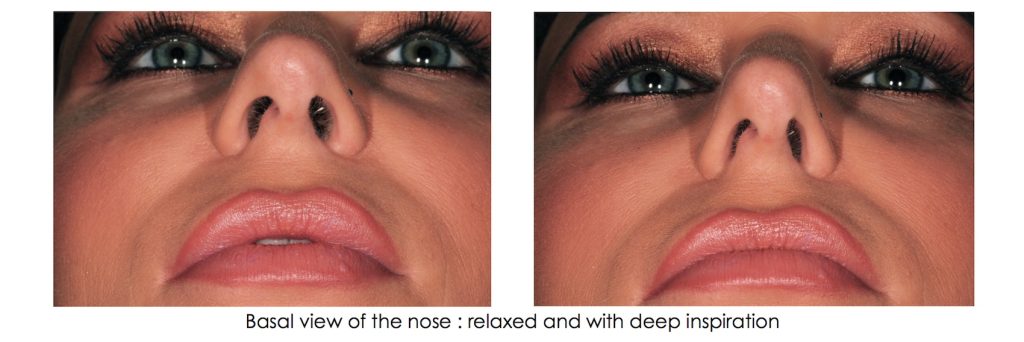
The basal view of the nose

Photograph the basal view of the nose when the patient is relaxed, and then when the patient breathes in deeply through their nose. Document whether the nasal valves collapse, restricting the airflow into the nasal cavity.
Photographic documentation is a great way to capture information and use it not only for record keeping, but to engage the patient in a guided discovery of their airway.
SPEAR ONLINE
Team Training to Empower Every Role
Spear Online encourages team alignment with role-specific CE video lessons and other resources that enable office managers, assistants and everyone in your practice to understand how they contribute to better patient care.

By: Robert Winter
Date: June 3, 2018
Featured Digest articles
Insights and advice from Spear Faculty and industry experts


