Caries Detection Tools: Select the Best for Your Practice
In Part I of this series of articles on caries detection, we examined the concept of dental caries, how we as clinicians may agree to disagree on what a radiographic image tells us, and when to treat.
In this article, we will examine the advancements in technology that exist on the market, enabling us a better chance to diagnose, restore or prevent further tooth destruction. For the sake of brevity, descriptions and information of the selected devices can be enhanced by clicking on the embedded links. The article is more of a primer for those interested in what exists on the market, older and newest. The price points of each of the electronic devices varied in quotes received and were omitted from this piece for that reason, as not to misinform our readers.
Caries Detector
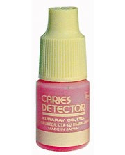
Caries detector, or disclosing solution, is not the oldest, but is of the simplest methods of detecting caries. It is widely used, cost effective and can well differentiate non-remineralizable from remineralizable dentin.
After drying and isolation of the area, place the staining solution for 10 seconds (while others suggest rinsing immediately, this is the manufacturer’s recommendation), rinse and observe areas of scarlet red (some manufacturers use a dark blue or green). This is the identified carious dentin that cannot be remineralized, and can be removed with spoon excavation and/or a round bur and slow speed handpiece. It can be especially helpful acting as a “flashlight” when excavating vast caries that run laterally and undermine buccal, lingual and palatal walls.
The use of the stain must be used with sound clinical judgement, using tactile feedback from an explorer and/or spoon excavator, as well as an understanding of mineralization zones of a tooth.
In an article/blog from Dr. Lee Ann Brady, she reminds us that “the challenge is that mineralization is not directly correlated with bacterial infiltration and caries. The circumpulpal dentin and that at the dentino-enamel junction is less mineralized. Therefore the dentin at the depth of the prep and close to the pulp often takes up the dye, resulting in a false positive result and potentially over preparation and increased risk of pulpal trauma.”1 Disclosing solution can be an effective tool in deciphering caries, and used to gain experience for all burgeoning dentists.
Microlux Transilluminator
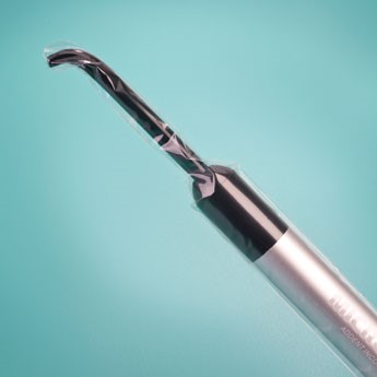
Shining a light through a tooth to diagnose maladies has been a consideration since the 1920s, when Cameron’s Dental Diagnostic Lamp was introduced by the Cameron Surgical Specialty Co.2 Today we have the Microlux Transilluminator from AdDent, Inc., a handheld wand-like device to detect interproximal caries, among other useful tools, such as oral cancer screenings, visualizing crown or root fractures, finding root canal orifices, and improving the visual field during apical surgeries.
Diagnodent
From KaVo, USA, comes a handheld laser device designed to aid in finding caries at their earliest stage. It measures reflected light, specifically laser fluorescence. Bacteria fluoresce when exposed to certain wavelengths of light. Diagnodent operates at 655 nm. It is at this wavelength that clean, healthy tooth structure exhibits little to no fluorescence, resulting in a very low reading on the device’s display.
Conversely, carious structures exhibit fluorescence relative to the degree of decay. The results are elevated readings on the display of the device. The clinician may remain focused on the teeth as a scaled audio signal sounds based on the severity of the caries/reading.

The teeth must be dried and a baseline must be recorded for each patient by selecting one healthy tooth. Record this in the chart to have for future readings. Click the link below for a video tutorial.
The Canary System
From Quantum Dental Technologies comes a laser-based handheld instrument featuring an integrated intraoral camera, detecting the presence of cracks and caries, before they are large enough to appear on radiographs. It is the only system that analyzes and measures the crystalline structures of a tooth. It can measure to a depth of 5 mm. It detects decay on all surfaces tooth surfaces including around margins of amalgams, resins, and crowns; smooth surfaces; pits/fissures; under sealants; and around orthodontic brackets.

When placed on the tooth surface, a low-powered, pulsating laser light is shone on the tooth during a three-second scan. The pulses of laser light generate photothermal (PTR) and luminescence (LUM) responses. By using a laser pulse at a frequency of 2 Hz, the laser light can penetrate below the tooth surface and permit detection of a carious lesion as small as 50 microns (20-times smaller than a millimeter) and as deep as 5 mm from the tooth surface.
For proximal caries diagnosis, the Canary System is more accurate than bitewing radiography. The Canary had a sensitivity of 92 percent compared to 67 percent for bitewing radiography, according to an independent human clinical study conducted at the University of Texas and released at the International Association for Dental Research Meeting in March 2015.3
CariVu
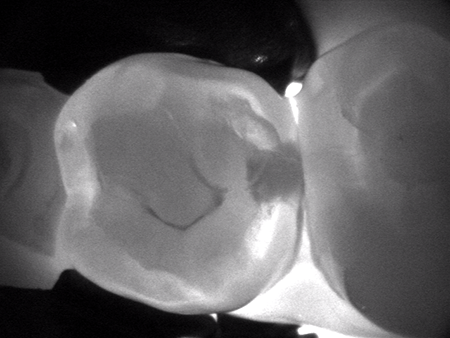
Dexis Dental has a new version of a handheld and portable transillumination device, CariVu. It uses patented technology to support the identification of occlusal, interproximal, and recurrent caries and cracks. The device is designed to engulf a tooth in their patented near infrared light. The tooth appears transparent. Carious lesions absorb light and try to mimic what interproximal decay looks like on a radiograph. Dexis believes they have an edge over devices that fluoresce in that the teeth do not need to be clean of bacteria, the device does not require calibration before each use, and the device reports more of the physical condition of a tooth.
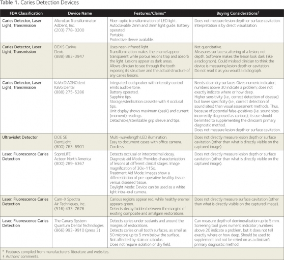
Below is a chart secured from an article for the ADA written by Dr. Douglas A. Young, professor, Department of Dental Practice, University of Pacific, and Dr. Rebecca Slayton, Chair of Pediatric Dentistry, University of Washington
Logicon Software 5.1
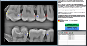
From Carestream Dental comes Logicon software, a computer-aided diagnostic tool. This is not a hand-held device. It uses a database of histologically validated caries cases to help determine whether caries are present.
According to Carestream, Logicon Software version 5.1 is clinically proven to help dentists find up to 20 percent more interproximal caries on permanent teeth than traditional methods, and is the only commercially available FDA-approved computer-aided radiographic caries diagnosis software. Latest software improvements further enhance the detection process. With a few clicks, Logicon Caries Detector PreScan applies the algorithm on all eligible inter-proximal surfaces within a radiograph to generate a view of existing caries, helping the dentist verify manual findings by focusing on surfaces that require further investigation.
Logicon software operates only within the confines of Carestream Dental Imaging software. It cannot be integrated with any other dental imaging software.
Conclusion
The intent of this article was to gather highlights so you may begin your own research for what may be the best fit for your practice in enhanced caries detection. There are no shortcuts in any diagnosis. Clinical visual, tactile, radiographic, patient dental history and susceptibility all play a role in individualized care. Experience is crucial. If you are short on experience, these tools can be of great value to use as an adjunct as well as educate. But even a veteran clinician can appreciate and enjoy these impressive diagnostics.
Who knows what is to come?
David St. Ledger, DDS, Visiting Faculty, Moderator, Contributing Author www.uppermontclairdental.com
References
1. Brady LA. Caries Detection Dye – Lee Ann Brady DMD. Lee Ann Brady DMDs Dental Blog 2011.
2. Transillumination: MedlinePlus Medical Encyclopedia. U.S National Library of Medicine 2015.
3. Uzamere EO, Jan J, Bakar WW, Mathews SM, Amaechi B. Clinical trial of the Canary System for proximal caries detection. International Association of Dental Research General Session 2015, Boston, MA
SPEAR campus
Hands-On Learning in Spear Workshops
With enhanced safety and sterilization measures in place, the Spear Campus is now reopened for hands-on clinical CE workshops. As you consider a trip to Scottsdale, please visit our campus page for more details, including information on instructors, CE curricula and dates that will work for your schedule.

By: Dave St. Ledger
Date: June 1, 2016
Featured Digest articles
Insights and advice from Spear Faculty and industry experts
