Single Anterior Implants Made Easy As 1-2-3
While restoring anterior teeth is a basic restorative procedure, creating an esthetic smile through restoration of the anterior teeth is a satisfying procedure for both the dentist and the patient.
One of the most challenging restorative procedures for dentists to perform is the replacement of a single anterior tooth. There are so many factors that play into these single restorations to re-create the beauty, form and function of the natural result. It can seem like an overwhelming task to achieve success in these situations. But in spite of these difficulties, replacement of the missing tooth through implant therapy can be a predictable and straightforward process.
Single anterior implant restorations are a mainstay in most dental practitioners’ “tool box.” Implants offer patients a wonderful alternative to fixed bridges and partial dentures. Through dental implant treatment, we are able to give patients confidence in mastication, stability of the bone and gingival tissues, and a beautiful smile by keeping a “one-tooth problem a one-tooth problem.”
Although many challenges and varying situations can interfere with an ideal result, following a logical sequence protocol and visualizing the outcome will provide a very predictable result.
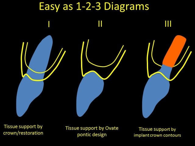
Easy As 1
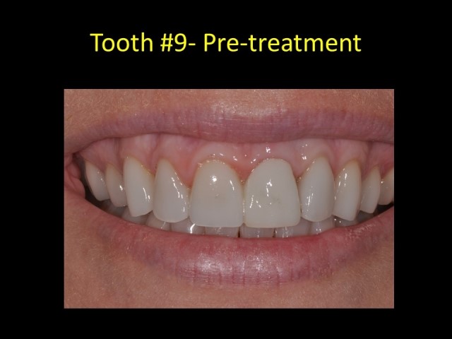
Tissue support for the gingival margins of a tooth comes from the tooth itself. The normal dentinogingival complex includes 1 mm of fibrous attachment, 1 mm junctional epithelium, and 1 mm sulcus depth. This is the “biologic width.”
In a healthy situation, the facial aspect of the natural tooth supports the facial margin. If a crown restoration or facial veneer were to be placed for esthetic or functional reasons, the restorative material would take the place of a natural tooth and be the “support substitute” for the facial profile to maintain the tissue support. Maintaining this support relationship is the “No. 1” parameter this article is referencing.
Esthetic dental restorations create natural contours and promote gingival health. Bulky or undercontoured restorations contribute to inflammation or lack of gingival support. Re-creating natural emergence profiles are paramount to esthetic appearance of the tooth and tissues.
Easy As 2
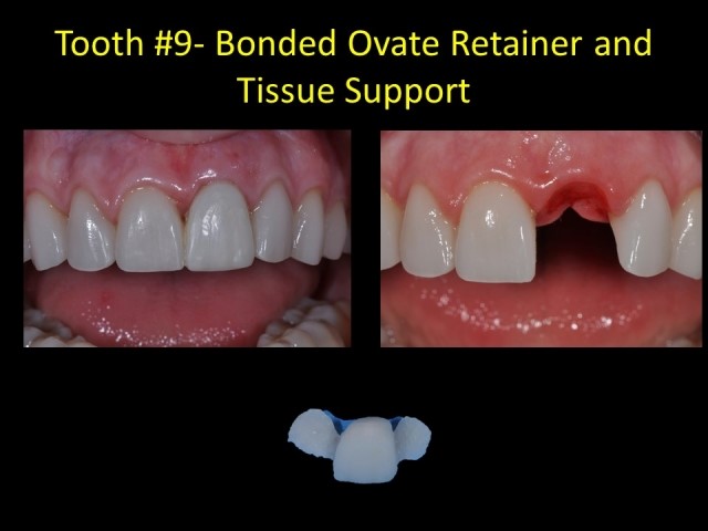
There are many situations when anterior teeth are lost or have to be removed. Loss of the tooth will result in loss of facial tissue support. Esthetics is compromised in those situations.
However, if the tooth replacement can substitute for this tissue support, esthetics, gingival health, and contour will be restored. The solution for this substitution lies in the understanding and application of ovate pontic design. The concepts for ovate pontics were developed in the early 1980s. This design was intended to create the appearance that the missing tooth was emerging from the gingival tissues as a natural tooth does. The “egg-shaped” emergent design provides support to the facial and interproximal tissues. The tissues hold their contours because of the hard structures that lend that support. By applying this ovate pontic technique to the interim replacement tooth (bonded retainer, “flipper,” etc.), a predictable facial margin may be maintained. The shape of the pontic will help “guide” the healing of the soft tissues.
The key to the ovate contour is to place the apex of the egg shape 2 mm apical and 2 mm lingual to the facial tissue margin. The emergence from the apex to the facial tissue margin provides the necessary support to maintain the height and contour of the facial margin. With this design, the gingival margins of the extracted tooth will be preserved and predictable health and esthetics will be achieved.
Easy As 3
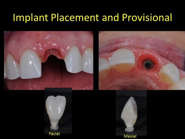
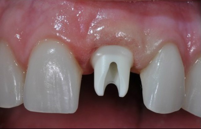
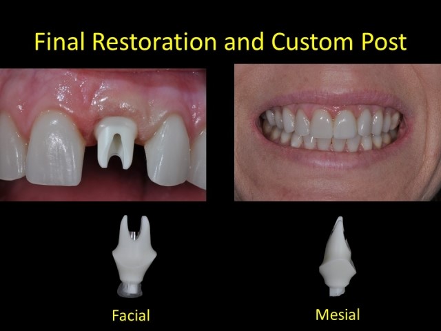
Once the extraction site is healed, a dental implant may be placed. By establishing and maintaining the gingival tissues through the ovate pontic design, the surgeon may utilize this facial margin as the reference point for the proper depth of the implant. Ideally, implant placement is 3 mm apical to the height of the facial tissue margin. This depth allows for adequate running room for contouring the custom provisional and, ultimately, the final post and crown. The facial contours of the provisional will mimic the gingival contours created by the pontic. Establishing this tissue reference point through proper pontic design and tissue scalloping results in an ideal outcome.
Visualizing the end in mind helps all members of the interdisciplinary team achieve predictable results. As can be seen from the diagrams, only three critical steps are necessary to attain predictable results. Maintaining support of the facial tissues with ideal crown emergence, ovate pontic design and properly contoured implant provisional provides the ideal results. Easy as 1-2-3!
SPEAR campus
Hands-On Learning in Spear Workshops
With enhanced safety and sterilization measures in place, the Spear Campus is now reopened for hands-on clinical CE workshops. As you consider a trip to Scottsdale, please visit our campus page for more details, including information on instructors, CE curricula and dates that will work for your schedule.

By: Jeffrey Bonk
Date: November 15, 2015
Featured Digest articles
Insights and advice from Spear Faculty and industry experts
