The Rule of Twos
If there were an observation that had the potential to prevent significant problems for our patients later in life, I am certain that dentistry (and YOU) would want to make that observation part of the standard of care. We ARE, after all, the prevention profession.
Still, change comes slowly and people change their ways and beliefs with resistance, a factor that science supports and encourages.1 Evidence must wind a tortuous trail to its acceptance as truth–and truth remains valid only until additonal evidence is put forth to replace it. To embrace science is to embrace skepticism and change, the skepticism comes easy.
Teeth: The Only Connection Between Occlusion and Peridontal Disease?
As a young dentist I read and heard that the only connection between occlusion and periodontal disease was the fact that both involved teeth. This observation was made in spite of the fact that much of the observation of the past had made significant connections between the two.2,3,4 The truth that came to be accepted, the lack of connection between the two, is being changed again as new evidence supports the observations of the past. 5,6 Today most periodontists would agree that treating traumatic occlusion is part of periodontal therapy. As is appropriate, not all share this belief and their skepticism will fuel the research that is necessary to firmly establish the evidence. There is a problem that now presents itself to the infomred clinician, how will you treat this patient with the knowledge of both opinions? I believe we must seek information and new evidence and act accordingly.

When it comes to growth and development, dentistry generally defers to the orthodontic community to provide standards that we can evaluate and refer our patients when therapeutic intervention is indicated. 7,8 Those of us with enough gray hair have heard just send them to me when they lose their baby teeth. We’ve also heard get that child to me as soon as you suspect they will need to know me for any reason whatsoever. Apparently there is controversy among orthodontists about when therapy should begin and whether or not influencing growth makes any real difference.9
It has been explained to me by some exceptional, caring clincians that the early intervention provides little more than a second chance to do orthodontics and that the second phase of care will still be performed with the same outcome had the first one been skipped. Feeling unqualified to enter into the argument, we generally do as requested and realize that each clinician is honestly doing what he or she feels to be in the best interest of the patient, doing what the truth as he or she knows it dictates their course.
TMJ Injury is Very Common in Children
I have always had an interest in tempero-mandibular disorders and in helping patients with pain feel better. That quest has led me to seek knowledge about the joints, the muscles, the nerves, the neuro-vasculature and the ways in which the dentition can contribute to the pain experienced by patients in these areas. That quest led me to seek greater understanding of a tempero-mandibular joint structure that is often compromised in patients with pain–the articular disk. It turns out that this compromise, a diskal displacement, is present in a lot of people– a lot of young people.10,11,12 Most of these patients do not have pain, rather they do not have the kind of pain that we equate with TMD because they are young and growing. The TMJ injury is usually caused by trauma to the face or whiplash type injuries, and there are a lot of them.13,14
Every parent has a story or two (or many) about the things they’ve seen happen to their child’s head and face – and remember, those are the times that they saw it happen. We know that the injuries occur, we know they injure joints and we know they cause growth changes and deficiences.15,16,17 The injuries also happen to adults to be sure, but the primary reason that we need to become adept at seeing the signs in children is that they still have growth potential and still have a chance to achieve normal growth if we can address the injury. Let me say that again. Discovered early enough in the growing child, many of these degenerative problems that we see in adults as TMD involving the joint can be successfully treated.
An injury to the growing TMJ that displaces a disk disrupts the growth center in the head of the condyle. Without the disk in place the condylar growth center becomes non-functional. This disruption creates an inability of the mandible to maintain its genetically programmed synergy with the maxilla and moves the patient toward a unilateral or bilateral Class II relationship. In the future, this displacement may also lead to degenerative reformation of the condyle, the articular emminence or both. Throughout these changes the teeth are trying to help. As the patient puts his or her teeth together and chews, squeezes or bruxes, the alveolar process tries to account for making things work.
Ask any orthodontist about their most difficult cases they experience and Class II patients are the first they mention.18,19 Is it any wonder that these can create a dilemma? The orthodontist is aiming for a moving target that is not moving the way he or she expects it to. Is it any surprise that there are significant relapses as things continue to reshape, reform, and often degenerate following the end of orthodontic therapy? As clinicians we need to become familiar with and comfortable with the signs that will prompt us to take early action. The sign that is most powerful is a change in the developing occlusion, bite drift.
Bad Joints Create Bad Bites
Bite drift is the observable change noted when the dimensions of the temporo-mandibular system are not growing synergistically (young people) or degenerating (adults). As dentists we have been trained to observe this and to act upon it. My favorite action for years was an appliance and an equilibration which seemed to work well for many people, but not for everyone. I am reasonably certain today that it worked on people who had a stable joint and failed on those who did not. Duh!
The problem is, I did not think to look at the foundation in a more critical way back then; I did not always image joints to see what foundation I was working with. My thought process was that bad bites create bad joints, and I acted accordingly. Today, I believe it’s predominantly bad joints that create bad bites. This means that a comprehensive evaluation of the foundation of the TMJ is critical to success. That means we must change our attitudes and our actions regarding imaging.
Imaging is indicated when we observe a loss in joint dimension presenting itself as bite drift. A developing Class II may be genetic, but if the patient has a normal maxilla and parents with normal maxillas and mandibles, it should raise some suspicions that this developing Class II may be of a non-genetic origin. Why are there so many Class II individuals with normal maxillas and normal parents? Maybe many of them are created from the failure of genetic potential to be realized. The loss of dimension leads back to the displacement of the disk and the changes in growth. Dimension lost with a displaced disk is 2mm to 3mm of vertical position and 2mm to 3mm of horizontal position as the condyle moves upward and backward into the area formerly occupied by the cartilage of the disk.20 Failure to develop and/or degeneration accounts for a change in condylar shape, ramus length and the drift toward Class II that continues.17,21
CBCT and MRI can provide a view into the TMJ that was not possible many years ago. Unfortunately, for many these are not utilized to the extent that would be helpful. CBCT raises the question of radiation exposure, certainly a question of momentous concern when dealing with a growing child. MRI is more difficult to obtain depending on your location and is generally expensive relative to other dental imaging that we are acquainted with. How can you know when to be patient and when to take action? The Rule of 2s22 is a good starting place.
The Rule of 2s
- Molar Class II by 2 mm
- Incisor horizontal uncouopling by 2 mm
- Incisor vertical uncoupling by 2 mm
- Combined molar shortening by 2 mm
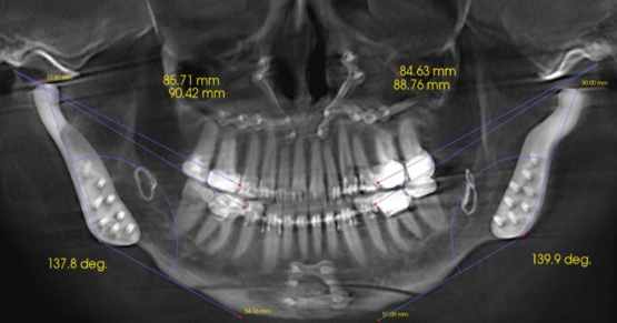
The reference position for these measurements is a fully seated condylar position. That position can be obtained using various methods. Bimanual Guidance (bilateral manipulation) works well with almost all children, a Leaf Gauge can be used if the patient can be comfortably loaded and a Lucia Jig can free the occlusion to get complete seating of the condyles. Use the method that lets you read the story the easiest. The fourth measurement is made on an image like a panorex, measuring the distance from the tympanic fissure to the mesial contact points of the maxilla first molar and the mandibular first molar. Subtracting the maxillary measurement from the mandibular measurement should result in a five (the maxillary first molar sits 5mm BACK onto the mandibular first molar). When this number is a three or less, there has been a drift to shorten the mandible relative to the maxilla, one of our warning signs from The Rule of 2s.
Comprehensive evaluation should consist of all of the signs and symptoms that our patients present with, and I believe that dentistry has created a stellar history of helping our patients see the things that we see. As we learn to see more, we have more to share. I never had a patient get angry with me for learning. The Rule of Twos is an observation that I believe we all need to add to our protocol, particularly for children. The evidence is there to my satisfaction that early detection can mean the difference between normal, comfortable, healthy and displaced, painful, diseased. Each of us would want that for our children. While you maintain your scientific skepticism, it’s important to stretch toward change and see what you find.
Want to know more?
E-mail me at gdewood@speareducation.com
References
- An Introduction to Scientific Research (McGraw-Hill, 1952), Bright W.
- The prevalence and possible role of nonworking contacts in periodontal disease. Periodontics. 1965 Sep-Oct;3(5):219-23.Yuodelis RA, Mann WV Jr.
- The angular bone defect and its relationship to trauma from occlusion and down growth of subgingival plaque. J Clin Periodontol. 1979 Apr;6(2):61-82. Waerhaug J.
- Tooth mobility and periodontal therapy. J Clin Periodontol. 1980 Jul 7(1):495-505. Fleszar J, Knowles J, Morrison E, Burgett F, Nissle R, Ramjford S.
- The effect of occlusal discrepancies on periodontitis. II. Relationship of occlusal treatment to the progression of periodontal disease. J Periodontol. 2001 Apr;72(4):495-505. Harrel SK1, Nunn ME.
- Longitudinal comparison of the periodontal status of patients with moderate to severe periodontal disease receiving no treatment, non-surgical treatment, and surgical treatment utilizing individual sites for analysis. J Periodontol. 2001 Nov;72(11):1509-19 Harrel SK, Nunn ME
- Guiding atypical facial growth back to normal. Part 2: Causative factors, patient assessment, and treatment planning. Int J Orthod Milwaukee. 2012 Spring;23(1):21-30. Galella SA1, Jones EB, Chow DW, Jones E 3rd, Masters A.
- Early treatment of Class II div 1 malocclusions. Orthod Fr. 2013 Mar;84(1):29-39. doi: 10.1051/orthodfr/2013037. Epub 2013 Mar 27. Chabre C.
- Early orthodontic treatment for growth modification by functional appliances–pros and cons. Refuat Hapeh Vehashinayim. 2014 Jan;31(1):25-31, 61.[ Tzemach M, Aizenbud D, Einy S.
- Disk displacement not present in 30 infants and young children. Oral Surg, Oral Med, Oral Path 87:15, 1999 Paesani et al:
- Asymptomatic disk displacement in 8 juveniles mean age 11. Am J Dentofacial Orthop 101:54, 1992 Hans et al:
- Asymptomatic disk displacement in 30% of adult volunteers. J Oral Max Surg 45:852, 1987 Kircos et al:
- Age and sex-related differences in 431 pediatric facial fractures at a level 1 trauma center. J Craniomaxillofac Surg. 2014 Apr 23. pii: S1010-5182(14)00114-0. doi: 10.1016/j.jcms.2014.04.002. [Epub ahead of print] Hoppe IC1, Kordahi AM2, Paik AM2, Lee ES2, Granick MS2.
- Youth Ice Hockey Injuries Over 16 Years at a Pediatric Trauma Center. Pediatrics. 2014 May 26. pii: peds.2013-3628. [Epub ahead of print] Polites SF, Sebastian AS, Habermann EB, Iqbal CW, Stuart MJ, Ishitani MB.
- Facial skeletal remodeling due to temporamandibular joint degeneration: An imaging study of 100 patients. AJR:155, 1990, 373-383. Schellas K, Piper M, Omlie D.
- Disorders of skeletal occlusion and tempero-mandibular joint disease. Northwest Dent 1989: Jan-Feb:35-42 Schellas Kp, Keck RJ
- Association between disk position and degenerative changes of the temporo-mandibular joints: an imaging study in subjects with TMD. The Journal of Craniomandibular Practice 2011:29(2):117-126. Sylvester D.
- Effectiveness of a fixed anterior bite plane in Class II deep-bite patients. Int J Orthod Milwaukee. 2014 Spring;25(1):15-20.Deregibus A, Debernardi CL, Persin L, Tugarin V, Markova M.
- Treatment of a complex malocclusion in a growing skeletal Class II patient. J Clin Orthod. 2014 Mar;48(3):181-9. El Refaei AK, Fayed MM, Heider AM, Mostafa YA.
- Magnetic resonance imaging in the evaluation of temporomandibular joint disc displacement-a review of 144 cases. Int J Oral Maxillofac Surg 2006;35:696-703. Whyte, AM; McNamara, D; Rosenberg, I; Whyte, AW.
- Condylar Shape in Relation to Anterior Disk Displacement in Juvenile Females. The Journal of Craniomandibular Practice 2011;29(2):100-110. Hasegawa, H.
- Piper, M. Personal conversation and notes
SPEAR STUDY CLUB
Join a Club and Unite with
Like-Minded Peers
In virtual meetings or in-person, Study Club encourages collaboration on exclusive, real-world cases supported by curriculum from the industry leader in dental CE. Find the club closest to you today!

By: Gary DeWood
Date: August 13, 2015
Featured Digest articles
Insights and advice from Spear Faculty and industry experts


