Positioning Maxillary Central Incisal Edges: Phonetics
In previous articles, I discussed the role of positioning the incisal edges of maxillary central incisors using primarily visual cues.

In this article I want to discuss a functional consideration when determining incisal edge position: phonetics. Any dentist who has done complete dentures, or who has restored anterior teeth, has experienced a patient who has returned with speech difficulties.
The key to understanding any changes in speech is to understand that the first thing necessary is to identify what “SOUND” the patient is struggling with. It’s also important to consider how much time the patient has had to adapt to the changes. The primary sounds that frequently change following alterations to anterior incisal edge position are: “F or V” sounds and “S” sounds.
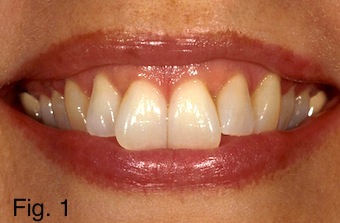
“F or V” sounds are the easiest to understand as they relate directly to the positioning of the maxillary incisal edges and don’t involve the mandibular anterior teeth. As most of us were trained, when a patient says “55,” the incisal edges of the anterior teeth should lightly contact the wet-dry line, (vermillion border), of the lower lip. The incisal edges impinging into the lip would be an example of excess incisal edge length. The female patient in Fig. 1 is an example of a patient who is tending toward a skeletal Class II relationship and has over erupted central incisors. In this photo she is saying “55.” It is easy to see that the incisal edges are impinging into the lip, indicating they are over erupted and positioned too far to the facial.
In my experience “S” sound problems are far more common than problems surrounding “F or V” sounds. I’ve found that “S” sound problems are also much more complicated. An “S” sound is the result of the maxillary anterior tooth position, mandibular anterior tooth position and the tongue interacting together. If a patient is having lisping problems following treatment, you need to assess how many teeth you altered to determine what is causing the problem. If you only altered the maxillary incisors, then the answer as to what produced the change is obvious, as it would be if you only treated the mandibular incisors.
Often a problem of lisping following treatment is produced because the anterior teeth are now making contact during the “S” sound. I would typically tell any patient that is having phonetic issues following treatment to wait at least four weeks to see if the issue resolves itself before making any major adjustments to incisal edge position.
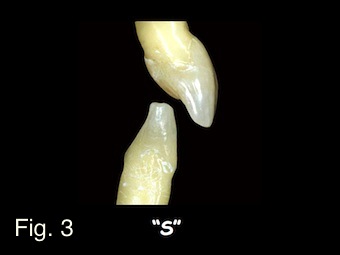
If at the end of four weeks the problem still exists, I would bring the patient in and ask them to say “66,” watching their anterior teeth as they make the sound. Roughly 70 percent of patients will make their “S” sounds by positioning the mandible so the incisors are end-to-end, Fig. 2. The remaining 30 percent make their “S” sound with the mandibular incisors somewhere along the lingual contour of the maxillary anterior teeth, Fig. 3. If the patient makes their “S” sound along the lingual of the anterior teeth as seen in Fig. 3, it means your maxillary incisal edge position is not the problem. The relationship of the lingual of the maxillary anterior teeth against the mandibular anterior incisal edges is the problem.
Regardless of where the patient makes their “S” sound, the correction is virtually always the same. Use some very thin articulating ribbon, I use “accufilm,” hold it against the maxillary incisors and ask the patient to say “66.” I then look for any areas of contact indicating an interference in the “closest speaking space,” you will now have to adjust the teeth until there is no tooth contact during the “S” sound. The challenge is deciding where to adjust, the upper or lower anterior teeth.
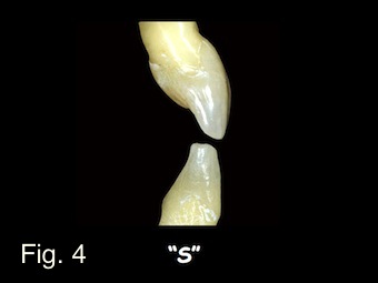
The patient in Fig. 4 is in full upper and lower provisional restorations, he is saying “66” you can see his anterior teeth are contacting and he has a lisp that hasn’t gone away after four weeks. The question is where to adjust. If I shorten the maxillary incisal edges I can eliminate the lisp, but may alter the esthetics. If I alter the mandibular anterior teeth by shortening them, I can eliminate the lisp and maintain the esthetics. However, now when he goes back into his intercuspal position he may have lost anterior occlusal contact, risking secondary eruption and the redevelopment of the lisp.
In most patients with end-to-end “S” positions, as seen in Fig. 4, I choose to alter the mandibular anterior teeth to correct speech but maintain esthetics, I then check the intercuspal position for anterior occlusal contact; if contact is still present you are done. If the contact has been removed, you can regain it by adding to the lingual of the maxillary anterior teeth which won’t affect the “S” sounds in the end-to-end position, so the lisp won’t return.
If your patient developed the lisp but their “S” position is the same as Fig. 3, the solution is almost always to alter the lingual of the maxillary anterior teeth which will eliminate the lisp, leave the incisal edge position intact and rarely removes the intercuspal position contact.
The bottom line is when you alter maxillary incisal edges you will eventually create some phonetic issues. Understanding these concepts can help solve the problems if providing the patient time to adapt doesn’t.
SPEAR campus
Hands-On Learning in Spear Workshops
With enhanced safety and sterilization measures in place, the Spear Campus is now reopened for hands-on clinical CE workshops. As you consider a trip to Scottsdale, please visit our campus page for more details, including information on instructors, CE curricula and dates that will work for your schedule.

By: Frank Spear
Date: October 24, 2013
Featured Digest articles
Insights and advice from Spear Faculty and industry experts

