- Beyond Restoration
- |
- Techniques / Materials
5 Reasons Why Dentists Should Be Using a Microscope

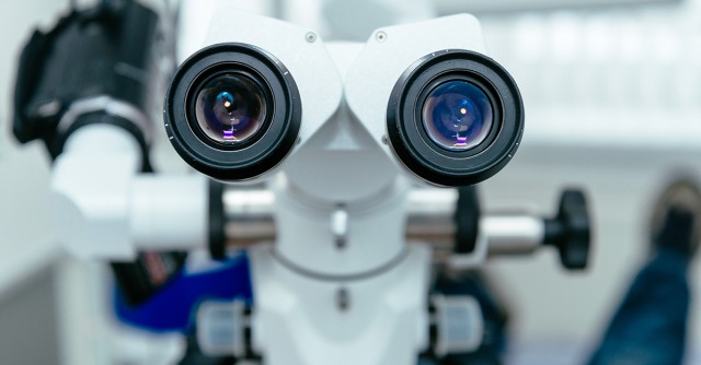
Magnification has revolutionized dentistry. Magnifying an image 2–4.5x with standard loupes is an asset, improving the operator’s visibility and, thereby, the ability to diagnose and provide treatment.
With magnification being the current standard of care in dentistry, why is it that the percentage of dentists using a microscope is still a minority? This article will discuss five advantages of implementing a microscope in your practice.
- Improved magnification. Remember when you started using loupes and could never practice without them because you knew what you weren’t seeing? That feeling holds when you go from using loupes to a microscope; wearing your loupes isn’t the same because you now know what you’re missing. Dental microscopes provide a range of magnification from 2.6x to 16x, with the average microscope dentist operating at an 8x magnification (compared with 2.5–4.5x magnification for most loupe users). This enhanced magnification can improve the accuracy of tooth preparations and margins, allow for more conservative treatment, and be kinder and gentler to adjacent teeth/restorations and the supporting soft tissues. Ultimately, higher magnification provides enhanced visibility in all aspects of dentistry, from diagnosing to prepping, seating, and finishing restorations.
- Improves ergonomics. Ergonomics is one of the biggest reasons dentists use a scope: It forces you to sit upright in your chair with improved posture. This fact alone can add years to your dental career by reducing strain on your neck and back. With correct positioning and posture, your body will feel better at the end of the day.
- Reduced eye fatigue/strain. The loupes technology uses “converging vision” because of the short working distance, which can cause eye fatigue and strain. The dental microscope functions differently: The higher magnification lens and distance from the operating field allow parallel vision of the working field, thereby reducing the strain and fatigue on the eyes.
- Improved lighting. The built-in light sources on the microscopes today are halogen, xenon, or LED. They allow excellent visibility and admission of light to areas in the mouth that are otherwise difficult to see, let alone access. Examples of this are common: deep interproximal decay on the mesial of second molars, looking down into a post space, finishing a composite restoration. The flood of light provided by a scope is a huge benefit in providing visibility to your working field without the hassle of cables or battery packs
- Documentation and education can be communicated to patients or other dentists. It can also be a good way to educate auxiliary staff.
There are many challenges to implementing a microscope into clinical practice: cost, staff acceptance, longer procedures initially, a steep learning curve, and most of all inconvenience. It’s not an easy thing to blend into your normal scheduled workday. That being said, the reality is, once you start using it and you experience the benefits, it’s tough to go back.
SPEAR ONLINE
Team Training to Empower Every Role
Spear Online encourages team alignment with role-specific CE video lessons and other resources that enable office managers, assistants and everyone in your practice to understand how they contribute to better patient care.

By: Greggory Kinzer
Date: August 9, 2016
Featured Digest articles
Insights and advice from Spear Faculty and industry experts


